 Old Web
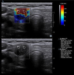
Transient elastography

Elastography is a medical imaging modality that maps the elastic properties and stiffness of soft tissue. The main idea is that whether the tissue is hard or soft will give diagnostic information about the presence or status of disease. For example, cancerous tumours will often be harder than the surrounding tissue, and diseased livers are stiffer than healthy ones. Elastography is a medical imaging modality that maps the elastic properties and stiffness of soft tissue. The main idea is that whether the tissue is hard or soft will give diagnostic information about the presence or status of disease. For example, cancerous tumours will often be harder than the surrounding tissue, and diseased livers are stiffer than healthy ones. The most prominent techniques use ultrasound or magnetic resonance imaging (MRI) to make both the stiffness map and an anatomical image for comparison. Elastography is used for the investigation of many disease conditions in many organs. It can be used for additional diagnostic information compared to a mere anatomical image, and it can be used to guide biopsies or, increasingly, replace them entirely. Biopsies are invasive and painful, presenting a risk of hemorrhage or infection, whereas elastography is completely noninvasive. Elastography is used to investigate disease in the liver. Liver stiffness is usually indicative of fibrosis or steatosis (fatty liver disease), which are in turn indicative of numerous disease conditions, including cirrhosis and hepatitis. Elastography is particularly advantageous in this case because when fibrosis is diffuse, a biopsy can easily miss sampling the diseased tissue, which results in a false negative misdiagnosis. Naturally, elastography sees use for organs and diseases where manual palpation was already widespread. Elastography is used for detection and diagnosis of breast, thyroid, and prostate cancers. Certain types of elastography are also suitable for musculoskeletal imaging, and they can determine the mechanical properties and state of muscles and tendons. Because elastography does not have the same limitations as manual palpation, it is being investigated in some areas for which there is no history of diagnosis with manual palpation. For example, magnetic resonance elastography is capable of assessing the stiffness of the brain, and there is a growing body of scientific literature on elastography in healthy and diseased brains. Preliminary reports on elastography used on transplanted kidneys to evaluate cortical fibrosis have been published showing promising results. Palpation is the practice of feeling the stiffness of a patient's tissues with the practitioner's hands. Manual palpation dates back at least to 1500 BC, with the Egyptian Ebers Papyrus and Edwin Smith Papyrus both giving instructions on diagnosis with palpation. In ancient Greece, Hippocrates gave instructions on many forms of diagnosis using palpation, including palpation of the breasts, wounds, bowels, ulcers, uterus, skin, and tumours. In the modern Western world, palpation became considered a respectable method of diagnosis in the 1930s. Since then, the practice of palpation has become widespread, and it is considered an effective method of detecting tumours and other pathologies. Manual palpation, however, suffers from several important limitations: it is limited to tissues accessible to the physician's hand, it is distorted by any intervening tissue, and it is qualitative but not quantitative. Elastography, the measurement of tissue stiffness, seeks to address these challenges.
- CCF Conference Analysis
- Map Galaxy
- Academic Report
- What's New