 Old Web
Obstetric ultrasonography

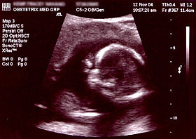
Obstetric ultrasonography is the use of medical ultrasonography in pregnancy, in which sound waves are used to create real-time visual images of the developing embryo or fetus in its mother's uterus (womb). The procedure is a standard part of prenatal care in many countries, as it can provide a variety of information about the health of the mother, the timing and progress of the pregnancy, and the health and development of the embryo or fetus.3D Ultrasound of fetal movements at 12 weeks75-mm fetus (about 14 weeks' gestational age)Fetus at 17 weeksFetus at 20 weeksContents in the cavity of the uterus seen at approximately 5 weeks of gestational age.Artificially colored, showing gestational sac, yolk sac and embryo (measuring 3 mm as the distance between the + signs).Embryo at 5 weeks and 1 day of gestational age (at top left) with discernible heartbeat.Embryo at 5 weeks and 5 days of gestational age with discernible heartbeat. Obstetric ultrasonography is the use of medical ultrasonography in pregnancy, in which sound waves are used to create real-time visual images of the developing embryo or fetus in its mother's uterus (womb). The procedure is a standard part of prenatal care in many countries, as it can provide a variety of information about the health of the mother, the timing and progress of the pregnancy, and the health and development of the embryo or fetus. The International Society of Ultrasound in Obstetrics and Gynecology (ISUOG) recommends that pregnant women have routine obstetric ultrasounds between 18 weeks' and 22 weeks' gestational age (the anatomy scan) in order to confirm pregnancy timing, to measure the fetus so that growth abnormalities can be recognized quickly later in pregnancy, and to assess for congenital malformations and multiple pregnancies (twins, etc). Additionally, the ISUOG recommends that pregnant women have obstetric ultrasounds between 11 weeks' and 13 weeks 6 days' gestational age in countries with resources to perform them (the nucal scan). Performing an ultrasound at this early stage of pregnancy can more accurately confirm the timing of the pregnancy and can also assess for multiple fetuses and major congenital abnormalities at an earlier stage. Research shows that routine obstetric ultrasound before 24 weeks' gestational age can significantly reduce the risk of failing to recognize multiple gestations and can improve pregnancy dating to reduce the risk of labor induction for post-dates pregnancy. There is no difference, however, in perinatal death or poor outcomes for babies. Below are useful terms on ultrasound: In normal state, each body tissue type, such as liver, spleen or kidney, has a unique echogenicity. Fortunately, gestational sac, yolk sac and embryo are surrounded by hyperechoic (brighter) body tissues. Traditional obstetric sonograms are done by placing a transducer on the abdomen of the pregnant woman. One variant, transvaginal sonography, is done with a probe placed in the woman's vagina. Transvaginal scans usually provide clearer pictures during early pregnancy and in obese women. Also used is Doppler sonography which detects the heartbeat of the fetus. Doppler sonography can be used to evaluate the pulsations in the fetal heart and bloods vessels for signs of abnormalities. Modern 3D ultrasound images provide greater detail for prenatal diagnosis than the older 2D ultrasound technology. While 3D is popular with parents desiring a prenatal photograph as a keepsake, both 2D and 3D are discouraged by the FDA for non-medical use, but there are no definitive studies linking ultrasound to any adverse medical effects. The following 3D ultrasound images were taken at different stages of pregnancy: A gestational sac can be reliably seen on transvaginal ultrasound by 5 weeks' gestational age (approximately 3 weeks after ovulation).The embryo should be seen by the time the gestational sac measures 20 mm, about five-and-a-half weeks. The heartbeat is usually seen on transvaginal ultrasound by the time the embryo measures 5 mm, but may not be visible until the embryo reaches 7 mm, around 7 weeks' gestational age. Coincidentally, most miscarriages also happen by 7 weeks' gestation. The rate of miscarriage, especially threatened miscarriage, drops significantly if normal heartbeat is detected. In the first trimester, a standard ultrasound examination typically includes: In the second trimester, a standard ultrasound exam typically includes:
- CCF Conference Analysis
- Map Galaxy
- Academic Report
- What's New