 Old Web
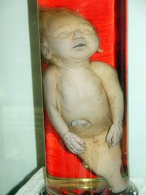
Sirenomelia

Sirenomelia, also called mermaid syndrome, is a rare congenital deformity in which the legs are fused together, giving the appearance of a mermaid's tail, hence the nickname. Sirenomelia, also called mermaid syndrome, is a rare congenital deformity in which the legs are fused together, giving the appearance of a mermaid's tail, hence the nickname. The word sirenomelia derives from the ancient Greek word seirēn, referring to the mythological Sirens, who were sometimes depicted as mermaids, and melos, meaning 'limb'. Sirenomelia is mainly characterized by the fusion of both legs with rotation of the fibula. It may include absence of the lower spine, abnormalities of the pelvis, renal organs, and was previously thought to be a severe form of sacral agensis/caudal regression syndrome, but more recently research verifies that these two conditions are not related. NORD has a separate report on caudal regression syndrome. In general, the more severe cases of limb fusion correlate with more severe dysplasia in the pelvis. Rather than the two iliac arteries present in fetuses with complete renal agenesis, fetuses with sirenomelia display no branching of the abdominal aorta, which is always absent. Associated defects recorded in cases of sirenomelia include neural tube defects (rachischisis, anencephaly, and spina bifida), holoprosencephaly, hypoplastic left heart syndrome, other heart defects, esophageal atresia, omphalocele, intestinal malrotation, persistent cloaca, and other limb defects (most commonly absence of the radius). Though obvious at birth, sirenomelia can be diagnosed as early as 14 weeks gestation on prenatal ultrasound. When there is low amniotic fluid around the fetus (oligohydramnios), the diagnosis is more difficult. The ultimate cause of sirenomelia is a subject of debate.The first hypothesis of its origin was developed in 1927 and proposed that a lack of blood supply to the lower limbs during their development is responsible for the defect. This 'vascular steal' hypothesis was developed in response to the observed absence or severe underdevelopment of the aorta below the umbilical artery, which 'steals' the blood supply from the lower limbs. Other hypotheses involve an insult to the embryo between 28–32 days affecting the caudal mesoderm, a teratogen exposure affecting the neural tube during neurulation, and a defect in the twinning process that either stops the process of caudal differentiation or generates a second primitive streak. Maternal diabetes mellitus has been associated with caudal regression syndrome and sirenomelia, although a few sources question this association. Prenatal cocaine exposure has also been suggested as an association with sirenomelia. In animal models, several genes have been found to cause or be associated with sirenomelia. The srn (siren) gene is observed to cause hindlimb fusion in homozygous mice. Mice with knockouts or mutations in both tsg1 and bmp7 will also have hindlimb fusion. Sirenomelia is classified by the skeletal structure of the lower limb, ranging from class I, where all bones are present and only the soft tissues are fused, to class VII where the only bone present is a fused femur.
- CCF Conference Analysis
- Map Galaxy
- Academic Report
- What's New