 Old Web
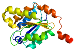
Gephyrin

1JLJ10243268566ENSG00000171723ENSMUSG00000047454Q9NQX3Q8BUV3NM_001024218NM_020806NM_145965NM_172952NP_001019389NP_065857NP_666077NP_766540Gephyrin is a protein that in humans is encoded by the GPHN gene.1ihc: X-ray Structure of Gephyrin N-terminal Domain1jlj: 1.6 Angstrom crystal structure of the human neuroreceptor anchoring and molybdenum cofactor biosynthesis protein gephyrin1t3e: Structural basis of dynamic glycine receptor clustering2fts: Crystal structure of the glycine receptor-gephyrin complex2fu3: Crystal structure of gephyrin E-domain Gephyrin is a protein that in humans is encoded by the GPHN gene. This gene encodes a neuronal assembly protein that anchors inhibitory neurotransmitter receptors to the postsynaptic cytoskeleton via high affinity binding to a receptor subunit domain and tubulin dimers. In nonneuronal tissues, the encoded protein is also required for molybdenum cofactor biosynthesis. Mutations in this gene may be associated with the neurological condition hyperekplexia and also lead to molybdenum cofactor deficiency. Numerous alternatively spliced transcript variants encoding different isoforms have been described; however, the full-length nature of all transcript variants is not currently known. The production of alternatively spliced variants is affected by noncoding regions within the gene. A ‘yin-yang’ noncoding sequence pair encompassing gephyrin has been identified. These sequences are opposites of each other - consisting of hundreds of divergent nucleotide states. Both of these patterns are uniquely human and evolved rapidly after splitting from their ancestral DNA pattern. The gephyrin yin and yang sequences are prevalent today in populations representing every major human ancestry. Gephyrin is a 93kDa multi-functional protein that is a component of the postsynaptic protein network of inhibitory synapses. It consists of 3 domains: N terminal G domain, C terminal E domain, and a large unstructured linker domain which connects the two. Although there are structures available for trimeric G and dimeric E domains, there is no structure available for the full length protein, which may be due to the large unstructured region which makes the protein hard to crystallize. But a recent study of the full length gephyrin by small-angle X-ray scattering shows that it predominantly forms trimers, and that because of its long linker region, it can exist in either a compact state or either of two extended states. Positive antibody staining for gephyrin at a synapse is most of the time consistent with the presence of glycine and/or GABAA receptors. Nevertheless, some exceptions can occur like in neurons of Dorsal Root Ganglions where gephyrin is absent despite the presence of GABAA receptors. Gephyrin is considered a major scaffolding protein at inhibitory synapses, analogous in its function to that of PSD-95 at glutamatergic synapses. Gephyrin was identified by its interaction with the glycine receptor, the main receptor protein of inhibitory synapses in the spinal cord and brainstem. In addition to its interaction with the glycine receptor, recent publications have shown that gephyrin also interacts with the intracellular loop between the transmembrane helices TM3 and TM4 of alpha and beta subunits of the GABAA receptor. Gephyrin displaces GABA receptors from the GABARAP/P130 complex, then brings the receptors to the synapse. Once at the synapse, the protein binds to collybistin and neuroligin 2. In cells, gephyrin appears to form oligomers of at least three subunits. Several splice variants have been described that prevent this oligomerization without influencing the affinity for receptors. They nevertheless affect the composition of inhibitory synapses and can even play a role in diseases like epilepsy. The gephyrin protein is also required during molybdenum cofactor assembly for insertion of molybdenum.
- CCF Conference Analysis
- Map Galaxy
- Academic Report
- What's New