 Old Web
Haptoglobin

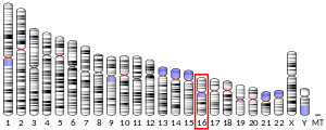
4X0L, 4WJG, 5HU6324015439ENSG00000257017ENSMUSG00000031722P00738Q61646NM_001126102NM_005143NM_001318138NM_017370NM_001329965NP_001119574NP_001305067NP_005134NP_001316894NP_059066Haptoglobin (abbreviated as Hp) is the protein that in humans is encoded by the HP gene. In blood plasma, haptoglobin binds to free hemoglobin, compared to hemopexin that binds to free heme, released from erythrocytes with high affinity and thereby inhibits its oxidative activity. The haptoglobin-hemoglobin complex will then be removed by the reticuloendothelial system (mostly the spleen). Haptoglobin (abbreviated as Hp) is the protein that in humans is encoded by the HP gene. In blood plasma, haptoglobin binds to free hemoglobin, compared to hemopexin that binds to free heme, released from erythrocytes with high affinity and thereby inhibits its oxidative activity. The haptoglobin-hemoglobin complex will then be removed by the reticuloendothelial system (mostly the spleen). In clinical settings, the haptoglobin assay is used to screen for and monitor intravascular hemolytic anemia. In intravascular hemolysis, free hemoglobin will be released into circulation and hence haptoglobin will bind the hemoglobin. This causes a decline in haptoglobin levels. This gene encodes a preproprotein that is processed to yield both alpha and beta chains, which subsequently combines as a tetramer to produce haptoglobin. Haptoglobin functions to bind free plasma hemoglobin, which allows degradative enzymes to gain access to the hemoglobin while at the same time preventing loss of iron through the kidneys and protecting the kidneys from damage by hemoglobin. For this reason, it is often referred to as the suicide protein. The cellular receptor target of Hp is the monocyte/macrophage scavenger receptor, CD163. Following Hb-Hp binding to CD163, cellular internalization of the complex leads to globin and heme metabolism, which is followed by adaptive changes in antioxidant and iron metabolism pathways and macrophage phenotype polarization. When Hb is released from RBCs within the physiologic range of Hp, the potential deleterious effects of Hb are prevented. However, during hyper-hemolytic conditions or with chronic hemolysis, Hp is depleted and Hb readily distributes to tissues where it might be exposed to oxidative conditions. In such conditions, heme can be released from ferric Hb. The free heme can then accelerate tissue damage by promoting peroxidative reactions and activation of inflammatory cascades. Hemopexin (Hx) is another plasma glycoprotein able to bind heme with high affinity. Hx sequesters heme in an inert, non-toxic form and transports it to the liver for catabolism and excretion. Haptoglobin is produced mostly by hepatic cells but also by other tissues such as skin, lung and kidney. In addition, the haptoglobin gene is expressed in murine and human adipose tissue. Haptoglobin had been shown to be expressed in adipose tissue of cattle as well. Haptoglobin, in its simplest form, consists of two alpha and two beta chains, connected by disulfide bridges. The chains originate from a common precursor protein, which is proteolytically cleaved during protein synthesis. Hp exists in two allelic forms in the human population, so-called Hp1 and Hp2, the latter one having arisen due to the partial duplication of Hp1 gene. Three genotypes of Hp, therefore, are found in humans: Hp1-1, Hp2-1, and Hp2-2. Hp of different genotypes have been shown to bind hemoglobin with different affinities, with Hp2-2 being the weakest binder.
- CCF Conference Analysis
- Map Galaxy
- Academic Report
- What's New