 Old Web
Intestinal schistosomiasis

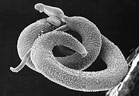
Schistosoma mansoni is a water-borne parasite of humans, and belongs to the group of blood flukes (Schistosoma). The adult lives in the blood vessels (mesenteric veins) near the human intestine. It causes intestinal schistosomiasis (similar to S. japonicum, S. mekongi, S. guineensis, and S. intercalatum). Clinical symptoms are caused by the eggs. As the leading cause of schistosomiasis in the world, it is the most prevalent parasite in humans. It is classified as a neglected tropical disease. As of 2016, 206.5 million people have schistosomiasis and S. mansoni is the major parasite. It is found in Africa, the Middle East, the Caribbean, Brazil, Venezuela and Suriname. Unlike other flukes (trematodes) in which sexes are not separate (monoecious), schistosomes are unique in that adults are divided into males and females, thus, (dioecious). However, the two adults live in permanent partnership, a condition called in copula; for this, they are considered as hermaphrodites. The life cycle of schistosomes includes two hosts: humans as definitive hosts, where the parasite undergoes sexual reproduction, and snails as intermediate hosts, where a series of asexual reproductive takes place. S. mansoni is transmitted through water, where freshwater snails of the genus Biomphalaria act as intermediate hosts. The larvae are able to live in water and infect the hosts by directly penetrating the skin. Prevention of infection is done by improved sanitation and killing the snails. Infection is treated with praziquantel. S. mansoni was first noted by Theodor Maximillian Bilharz in Egypt in 1851, while discovering S. haematobium. Sir Patrick Manson identified it as unique species in 1902. Louis Westenra Sambon gave the name Schistosomum mansoni in 1907 in honour of Manson. Schistosomes, unlike other trematodes, are long and cylindrical worms. The male S. mansoni is approximately 1 cm long (0.6–1.1 cm) and is 0.1 cm wide. It is white, and it has a funnel-shaped oral sucker at its anterior end followed by a second pediculated ventral sucker. The external part of the worm is composed of a double bilayer, which is continuously renewed as the outer layer, known as the membranocalyx, and is shed continuously. The tegument bears a large number of small tubercules. The suckers have small thorns in their inner part as well as in the buttons around them. The male genital apparatus is composed of 6 to 9 testicular masses, situated dorsally. There is one deferent canal beginning at each testicle, which is connected to a single deferent that dilates into a reservatory, the seminal vesicle, located at the beginning of the gynaecophoric canal. The copula happens through the coaptation of the male and female genital orifices. The female has a cylindrical body, longer and thinner than the male's (1.2 to 1.6 cm long by 0.016 cm wide). It has the general appearance of a roundworm. The female parasite is darker, and it looks gray. The darker color is due to the presence of a pigment (hemozoin) in its digestive tube. This pigment is derived from the digestion of blood. The ovary is elongated and slightly lobulated and is located on the anterior half of the body. A short oviduct conducts to the ootype, which continues with the uterine tube. In this tube it is possible to find 1 to 2 eggs (rarely 3 to 4) but only 1 egg is observed in the ootype at any one time. The genital pore opens ventrally. The posterior two-thirds of the body contain the vittelogenic glands and their winding canal, which unites with the oviduct a little before it reaches the ootype. The digestive tube begins at the anterior extremity of the worm, at the bottom of the oral sucker. The digestive tube is composed of an esophagus, which divides in two branches (right and left) and that reunite in a single cecum. The intestines end blindly, meaning that there is no anus. The eggs are oval-shaped, measuring 115-175 µm long and 45-47 µm wide, and ~150 µm diameter on average. They have pointed spines towards the broader base on one side, i.e. lateral spines. This is an important diagnostic tool because co-infection with S. haematobium (having a terminal-spined eggs) is common, and they are hard to distinguish. When the eggs are released into the water, a lot of them are immature and unfertilised so that they do not hatch. When the eggs are larger than 160 µm in diameter, they also fail to hatch. The miracidium (from the Greek word μειράκιον, meirakion, meaning youth) is pear-shaped, and gradually elongates as it ages. It measures about 136 μm long and 55 μm wide. The body is covered by anucleate epidermal plates separated by epidermal ridges. The epidermal cells give off numerous hair-like cilia on the body surface. There are 17-22 epidermal cells. Epidermal plate is absent only at the extreme anterior called apical papilla, or terebratorium, which contains numerous sensory organelles. Its internal body is almost fully filled with glycogen particles and vesicles.
- CCF Conference Analysis
- Map Galaxy
- Academic Report
- What's New