 Old Web
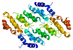
TRPV2

2F375139322368ENSG00000187688ENSMUSG00000018507Q9Y5S1Q9WTR1NM_016113NM_011706NP_057197NP_035836Transient receptor potential cation channel subfamily V member 2 is a protein that in humans is encoded by the TRPV2 gene. TRPV2 is a nonspecific cation channel that is a part of the TRP channel family. This channel allows the cell to communicate with its extracellular environment through the transfer of ions, and responds to noxious temperatures greater than 52 °C. It has a structure similar to that of potassium channels, and has similar functions throughout multiple species; recent research has also shown multiple interactions in the human body.2f37: Crystal structure of the ankyrin repeat domain of human TRPV2 Transient receptor potential cation channel subfamily V member 2 is a protein that in humans is encoded by the TRPV2 gene. TRPV2 is a nonspecific cation channel that is a part of the TRP channel family. This channel allows the cell to communicate with its extracellular environment through the transfer of ions, and responds to noxious temperatures greater than 52 °C. It has a structure similar to that of potassium channels, and has similar functions throughout multiple species; recent research has also shown multiple interactions in the human body. The vanilloid TRP subfamily (TRPV) named after the vanilloid receptor 1 consist of six members, four of them (TRPV1-TRPV4) have been related to thermal sensation. TRPV2 shares 50% of its homology with TRPV1. Compared to TRPV1 channels, TRPV2 channels do not open in response to vanilloids like capsaicin or thermal stimuli around 43 °C. This may be due to the composition of the ankyrin repeat domains in TRPV2, which are different than those in TRPV1. However, TRPV2 channels can open by noxious temperatures greater than 52 °C. TRPV2 initially was characterized as a noxious heat sensor channel, but more evidence suggest its importance in various osmosensory and mechanosensory mechanisms. The channel can open in response to a variety of stimuli including hormones, growth factors, mechanical stretching, heat, osmotic swelling, lysophospholipids, and cannabinoids. These channels are expressed in medium to large diameter neurons, motor neurons, and other non-neuronal tissues like the heart and lungs, which indicates its versatile function. The channel has an important role for basic cell function including contraction, cell proliferation, and cell death. The same channel can have different functions depending on the type of tissue. Other roles of TRPV2 continue to be explored in an attempt to define the role of translocation of TRPV2 by growth factors. TRPV2 was independently discovered by two research groups and described in 1999. It was identified in the lab of David Julius as a close homolog of TRPV1, known as the first identified thermosensitive ion channel. Itaru Kojima from Gunma University was looking for a protein which is responsible for the entry of calcium into cells in response to insulin-like growth factor-1 (IGF-1). Upon stimulation of cells with IGF-1, it was discovered that TRPV2 translocates towards and integrates into the cell membrane and increases intracellular calcium concentrations. TRPV2 channel has a similar structure to potassium channels, which are the largest ion channel family. This channel is composed of six transmembrane spanning regions (S1-S6) with a pore forming loop between S5 and S6. The pore forming loop also defines the selectivity filter, which determines the ions that are able to enter the channel. The S1-S4 region, as well as the N and C terminals of the protein, is important in reference to the gating of the channel. Although TRPV2 is a nonspecific cation channel, it is more permeable to calcium ions; calcium is an intracellular messenger and plays a very important role in a variety of different cellular processes. At rest, the pore channel is closed; in the activated state, the channel opens, allowing the influx of sodium and calcium ions that initiates an action potential. The TRPV subfamily of channels of 1 through 4 have unique functions. One important variation is that these channels trigger cellular signaling pathways via non-selective cation flux, making them unique. Specifically, the TRPV2 channel has structural similarities amongst the other members of the TRPV family. For instance, the channel consists of six transmembrane domains and a pore forming loop between S5 and S6. Within the human genome, putative homologs can be found. This suggests that the amino acids and proteins coded come from a common ancestor where their structures are conserved in function. Among the subfamily, TRPV2 and TRPV1 share 50% of their sequence identity not only in humans, but in rats as well. The rat TRPV2 can be comparable to that of humans because they exhibit similar surface localization among one another. Each channel possesses ATP binding regions and the 50% sequence identity between TRPV1 and TRPV2 suggests that both channel’s Ankyrin repeat domain (ARD) bind to different regulatory ligands as well. The channels structure can be observed as similar to that of potassium channels. In knockout mice, the physiological thermal responses show similar activation to wild-type mice. On top of that, humans, rats, and mice are considered orthologues. In homo sapiens, there is broad expression of TRPV2 in the lymph nodes, spleen, lung, appendix, and placenta; it is mostly expressed in the lungs. TRPV2 is majorly in a subpopulation of medium to large sensory neurons, as well as being distributed in the brain and spinal cord. The mRNA expression of TRPV2 is also found in human pulmonary and umbilical vein endothelial cells. Based on mRNA expression of TRPV2 in mice, it is also speculated that it is expressed in arterial muscle cells, which can then be influenced by blood pressure; though it was evident that TRPV2 expression was localized in the intracellular area, some growth factors localized it to the plasma cell membrane. In circulatory organs, studies and data suggest that TRPV2 may be a mechanosensor, meaning that it can sense changes in external stimuli; the mechanisms involved in opening TRPV2 by membrane stretching or hypoosmotic cell swelling have not yet been determined. In mus musculus (house mouse), TRPV2 functions as a protein coding gene. There is broad expression of TRPV2 in the thymus, placenta, cerebellum, and spleen; it is most commonly expressed in the thymus. The thymus is a lymphoid organ involved in the function of the immune system, where T cells mature. T cells are an important component to the adaptive immune system, because it is where the body adapts to foreign substances; this demonstrates TRPV2’s importance in the immune system. TRPV2 in mus musculus is also activated by hypo-osmolarity and cell stretching, indicating that TRPV2 plays a role in mechanotransduction in mice as well. In experiments with knockout mice (TRPV2KO mice), it was found that TRPV2 is expressed in brown adipocytes and in brown adipose tissue (BAT). It can be concluded that TRPV2 plays a role in BAT thermogenesis in mice, since it was found that a lack of TRPV2 impairs this thermogenesis in BAT; given these results, this could be a target for human obesity therapy. In rattus norvegicus (Norway rat), there is broad expression of TRPV2 in the adrenal glands and the lungs, being most present in the adrenal glands. TRPV2 is also present in the thymus and spleen, but not in high amounts. Without using any external growth factors, TRPV2 is highly specific to the plasma cell membrane in rat adult dorsal root ganglions, cerebral cortex, and arterial muscle cells.
- CCF Conference Analysis
- Map Galaxy
- Academic Report
- What's New