 Old Web
Omphalocele

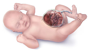
Omphalocele, also called exomphalos, is a rare abdominal wall defect in which the intestines, liver and occasionally other organs remain outside of the abdomen in a sac because of failure of the normal return of intestines and other contents back to the abdominal cavity during around the ninth week of intrauterine development. Omphalocele, also called exomphalos, is a rare abdominal wall defect in which the intestines, liver and occasionally other organs remain outside of the abdomen in a sac because of failure of the normal return of intestines and other contents back to the abdominal cavity during around the ninth week of intrauterine development. Omphalocele occurs in 1 in 4,000 births and is associated with a high rate of mortality (25%) and severe malformations, such as cardiac anomalies (50%), neural tube defect (40%), exstrophy of the bladder and Beckwith–Wiedemann syndrome. Approximately 15% of live-born infants with omphalocele have chromosomal abnormalities. About 30% of infants with an omphalocele have other congenital abnormalities. The sac, which is formed from an outpouching of the peritoneum, protrudes in the midline, through the umbilicus (navel). It is normal for the intestines to protrude from the abdomen, into the umbilical cord, until about the tenth week of pregnancy, after which they return to inside the fetal abdomen. The omphalocele can be mild, with only a small loop of intestines present outside the abdomen, or severe, containing most of the abdominal organs. In severe cases surgical treatment is made more difficult because the infant's abdomen is abnormally small, having had no need to expand to accommodate the developing organs. Larger omphaloceles are associated with a higher risk of cardiac defects. Omphalocele is caused by malrotation of the bowels while returning to the abdomen during development. Some cases of omphalocele are believed to be due to an underlying genetic disorder, such as Edward's syndrome (trisomy 18) or Patau syndrome (trisomy 13). Beckwith–Wiedemann syndrome is also associated with omphaloceles. Exomphalos is caused by a failure of the ventral body wall to form and close the naturally occurring umbilical hernia that occurs during embryonic folding which is a process of embryogenesis. The normal process of embryogenesis is that at 2 weeks gestation the human embryo is a flat disc that consists of three layers, the outer ectoderm and inner endoderm separated by a middle layer called the mesoderm. The ectoderm gives rise to skin and the CNS, the mesoderm gives rise to muscle and the endoderm gives rise to organs. The focus areas for exomphalos are that the ectoderm will form the umbilical ring, the mesoderm will form the abdominal muscles and the endoderm will form the gut. After the disc becomes tri-layered, it undergoes growth and folding to transform it from disc to cylinder shaped. The layer of ectoderm and mesoderm in the dorsal axis grow ventrally to meet at the midline. Simultaneously, the cephalic (head) and caudal (tail) ends of these layers of the disc fold ventrally to meet the lateral folds in the center. The meeting of both axis at the center form the umbilical ring. Meanwhile, the endoderm migrates to the center of this cylinder.
- CCF Conference Analysis
- Map Galaxy
- Academic Report
- What's New