 Old Web
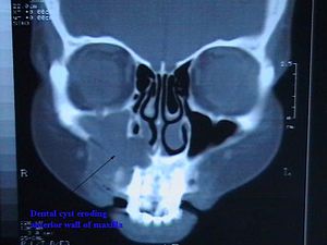
Periapical cyst

The periapical cyst is the most common odontogenic cyst. Periapical is defined as 'the tissues surrounding the apex of the root of a tooth' and a cyst is 'a pathological cavity lined by epithelium, having fluid or gaseous content that is not created by the accumulation of pus.' Most frequently located in the maxillary anterior region, it is caused by pulpal necrosis secondary to dental caries or trauma. The cyst has lining that is derived from the epithelial cell rests of Malassez which proliferate to form the cyst. Highly common in the oral cavity, the periapical cyst is asymptomatic, but highly significant because a secondary infection can cause pain and damage. In radiographs, it appears a radiolucency (dark area) around the apex of a tooth's root. The periapical cyst is the most common odontogenic cyst. Periapical is defined as 'the tissues surrounding the apex of the root of a tooth' and a cyst is 'a pathological cavity lined by epithelium, having fluid or gaseous content that is not created by the accumulation of pus.' Most frequently located in the maxillary anterior region, it is caused by pulpal necrosis secondary to dental caries or trauma. The cyst has lining that is derived from the epithelial cell rests of Malassez which proliferate to form the cyst. Highly common in the oral cavity, the periapical cyst is asymptomatic, but highly significant because a secondary infection can cause pain and damage. In radiographs, it appears a radiolucency (dark area) around the apex of a tooth's root. Secondary symptoms of periapical cysts include inflammation and infection of the pulp causing dental caries. This infection is what causes necrosis of the pulp. Expansion of the cyst causes erosion of the floor of the maxillary sinus. As soon as it enters the maxillary antrum, the expansion rate increases due to available space for expansion. Performing a percussion test by tapping the affected teeth will cause shooting pain. This is often clinically diagnostic of pulpal infection. Radiographically, it is virtually impossible to differentiate granuloma from a cyst. If the lesion is large it is more likely to be a cyst. Radiographically, both granulomas and cysts appear radiolucent. Many lesions of the mandible in particular appear cystlike in appearance. It is often necessary to obtain a biopsy and evaluate the tissue under a microscope to accurately identify the lesion.
- CCF Conference Analysis
- Map Galaxy
- Academic Report
- What's New